Introduction
Pro-life issues are among the most important facing Catholics and other believers throughout the world. Abortion must certainly rank as the top issue in the pro-life agenda. Even among non-Christians, polls reveal that between 15% and 75% of persons believe abortion should in illegal in all or most cases.[1]
In 2019 alone, about 889,000 abortions took place in the United States;[2] about 86,000 in Canada,[3] and about 42.3 million worldwide.[4]
Within the Catholic Tradition an abortion is considered the direct killing of the innocent, which is always considered to be a moral wrong. For pro-life advocates it is the major issue in their efforts to save innocent lives.
Embryology is the scientific study of the process by which an organism develops from a single cell to a fetus. Being a science, there is a myriad of technical terms describing the various stages of development. Purposeful misuse of language and terminology can sometimes be used to sway people as to what may constitute an abortion. It is common to hear proponents of abortion call an embryo a “mass of cells”, or “tissue that is not yet a person”.
Reproduction is not a moment, but rather a process. It is the purpose of this paper to serve as a primer on human embryology so as to assist with an understanding of the process. It is hoped that this would aid in not only being misled by language used to defend abortion, but also to become a more capable defender of the child-in-utero. As an example, some may believe that the term pre-embryo is sometimes used to describe that the organism is not yet human. This is not the case. As we review the process it will become clear that the scientific term pre-embryo is used to describe the fertilized egg up to 14 days old, at which time it becomes implanted in the uterus.
The Catholic Church teaches that a human life exists from the moment of fertilization.[5] Language cannot change that pronouncement. This must serve as the bedrock for the remainder of this article. The term pre-embryo is not in conflict with Catholic teaching. It is simply a scientific term describing one of the various stages from the beginning of pregnancy to the birth of a child.
As previously noted, human embryology is the scientific study of the development of the person through the various stages from the fertilization of the egg to that of fetal development and subsequent birth.
The Embryology of the Human Fetus
The process of reproduction in humans requires specialized cells known as gametes. In the male, the gamete is known as the sperm, while in the female it is called the ovum. The sperm may also be known as a spermatozoa, and the ovum as an oocyte or egg.
The gametes are produced in the male testis and the female ovary by a complicated type of cell division known as meiosis. This process results in gametes which are unique in that they are haploid cells, meaning that they contain only half of the genetic material, known as chromosomes, as all the other cells in the body.[6]
Human development begins at the time of fertilization of the woman’s oocyte by the man’s sperm.
During the first 24 hours after intercourse, by a process called capacitation, the sperm becomes able to penetrate the oocyte. After the sperm enters the egg various hormonal changes occur which prevent other sperm from entering it. Then by a process called syngamy, the two haploid nuclei (one from the egg and one from the sperm), fuse into a diploid nucleus within the now fertilized egg or zygote. Now the fertilized egg carries the full chromosome set for a human person.
During this time frame, twinning may occur. Monozygotic twins are known as identical twins. This occurs when a zygote (fertilized egg) splits and each develops into an embryo. Dizygotic twins (fraternal twins) occur when an ovary releases two eggs at about the same time and both are fertilized by separate sperm cells.
Over the next 14 days the zygote begins a series of rapid cell divisions by which one cells splits into two cells, two cells into four cells, and so forth. The 16-cell stage is known as the morula. Subsequently, after more cell divisions, the blastocyst develops with cells already beginning to differentiate into various tissue types. The result is a pre-embryo that is a unique entity, the product of both the male and female parents.
At about the 14th day, the pre-embryo reaches the uterus where it fuses itself to the wall of the uterus. At this time, a highly complex process begins by which there is rapid cell division into two different types of cells: embryoblast cells and trophoblast cells. The embryoblasts will continue to divide into what will eventually become the embryo and subsequently the baby. The trophoblasts will continue to divide and ultimately form the placenta. The blastocyst is now beginning to differentiate into the various organ tissues of the baby. This is now the embryo stage.
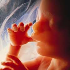
By three weeks, the placenta provides a rich source of nutrients for the developing baby which at this time is less than an eighth of an inch in size. By one month after fertilization, tissues have differentiated enough that the heart begins to form along with the circulatory system. The nervous tissue of the spine can also be identified. By ten weeks the fetus begins to resemble a human. By fourteen weeks all the major characteristics of a human person have formed. From this point in time until birth there is primarily growth in size of the fetus (now often called the baby) rather than any significant further development.
Eventually, because of the rapid growth up until the ninth month, the placenta can no longer provide adequate nutrition for the growing baby. The hormone oxytocin is released which stimulates the uterus to contract and begins the process of labor.
GLOSSARY
Blastocyst – an early stage of fetal development occurring at about the time of implantation into the uterus. It is defined by a hollow cavity within the developing mass of cells.
Capacitation – the process by which a sperm cell becomes capable of penetrating and entering the oocyte (egg).
Chromosomes – the genetic material within the cells.
Diploid – a cell which contains the full complement of genetic material (chromosomes).
Dizygotic – twins who develop from two separate zygotes (fraternal).
Egg – the female gamete. Also known as the oocyte or the ovum.
Embryo – the organism during the early stages of development, generally to about 8-10 weeks.
Embryoblast – differentiated cells that will eventually develop into the embryo.
Embryology – the study of the various stages of development from fertilization until birth.
Fertilization – the process of sexual reproduction by which the union of two haploid cells (gametes) forms a diploid cell (zygote).
Fraternal – twins who develop from two separate zygotes (dizygotic).
Gametes – cells which contain half of the genetic material (sperm and oocytes).
Haploid – cells which contain the full complement of genetic material (zygotes).
Meiosis – a complex process by which haploid cells are formed from diploid cells.
Monozygotic – identical twins developing from a single zygote.
Morula – the 16-cell stage of development after fertilization.
Oocyte – the female germ cell.
Ovary – the female reproductive organ in which eggs are formed.
Ovum – the female germ cell.
Oxytocin – the hormone that stimulates the uterus to contract and for labor to begin.
Placenta – the organ within the uterus which provides essential nourishment for the developing embryo/fetus.
Pre-embryo – an early stage of fetal development after fertilization is complete until about implantation in the uterus.
Sperm – the male germ cell.
Spermatozoa – the male germ cell.
Syngamy – the process by which two haploid cells merge into a single diploid cell.
Testes – the male reproductive organ in which sperm are formed.
Trophoblast – differentiated cells that will eventually develop into the placenta.
Zygote – the fertilized egg.
NOTES
A comprehensive review of human embryology can be found at: https://embryology.med.unsw.edu.au/embryology/index.php/Embryonic_Development
REFERENCES
[1] Pew Research Center. 2014. “Religious Landscape Study.” Accessed August 3, 2021. https://www.pewforum.org/religious-landscape-study/views-about-abortion/
[2] Abort73.com. 2020. “U.S. Abortion Statistics.” Accessed August 3, 2021. https://abort73.com/abortion_facts/us_abortion_statistics/
[3] Abortion Rights Coalition of Canada. 2021. “Statistics – Abortion in Canada.” Accessed August 3, 2021. https://www.arcc-cdac.ca/wp-content/uploads/2020/07/statistics-abortion-in-canada.pdf
[4] Decision Magazine. 2020. “Abortion Leading Cause of Death Worldwide in 2019.” Accessed August 3, 2021. https://decisionmagazine.com/abortion-leading-cause-death-worldwide-2019/
[5] In Donum Vitae (1987), the Congregation for the Doctrine of the Faith stated: “From the moment of conception, the life of every human being is to be respected in an absolute way because man is the only creature on earth that God has wished for himself and the spiritual soul of each man is ‘immediately created’ by God; his whole being bears the image of the Creator.” Subsequently in 1997, the Pontifical Academy for Life states: From a biological standpoint, the formation and the development of the human embryo appears as a continuous, coordinated and gradual process from the time of fertilization, at which time a new human organism is constituted, endowed with the intrinsic capacity to develop by himself into a human adult.
[6] Biology Online. 2021. “meiosis.” Accessed August 3, 2021. https://www.biologyonline.com/dictionary/meiosis


